
A healthy, 25-year-old woman presented to the emergency department complaining of abdominal pain and a low-grade fever for 5 days. A physical examination demonstrated a temperature of 37.8 °C and tenderness in the right lower quadrant. Blood test results included a white blood cell count of 13,000 cells/μL and a C-reactive protein level of 5 mg/dL. The emergency physician performed a point-of-care ultrasound examination, which demonstrated the marked thickening (up to 8 mm) of the terminal ileum adjacent to the ileocecal valve. The appendix had a normal diameter of 6 mm.
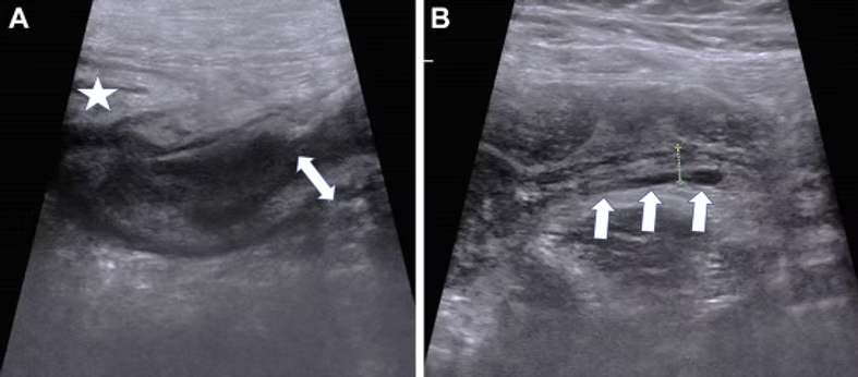
A, Longitudinal abdominal ultrasonographic view of the thickened (up to 8 mm) walls of the terminal ileum (double-sided arrow), adjacent to the thickened wall of the cecum (star). B, Longitudinal abdominal ultrasonographic view of the full-length, normal appendix (white arrows).
Read the full publication:
https://www.annemergmed.com/article/S0196-0644(22)00319-5/fulltext