
The use of sonography for diagnosing inflammatory bowel disease (IBD) has been reported in the radiology literature but is not common practice in the hands of emergency physicians (EPs). We present a series of three cases where IBD was managed by an EP using point-of-care ultrasonography (POCUS) and discuss the sonographic features of IBD, including bowel wall thickening, increased blood flow on color Doppler, infiltration of surrounding fatty tissue, and the presence of intraperitoneal fluid. Complications such as bowel strictures and peri-colic abscesses are also described. We suggest that the use of POCUS for the assessment of IBD patients in the ED may expedite both diagnosis and treatment, as well as minimize the use of additional imaging.
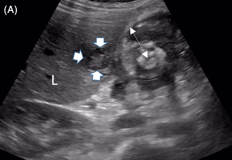
A, Transverse sonogram of the hepatic flexure demonstrating a narrowed lumen, significant thickening of the bowel wall (double arrow), and heterogeneous echogenicities in the bowel wall, reflecting inflammatory changes. There is an adjacent hypoechoic, heterogeneous liver lesion suspicious for an abscess (arrows). L, liver
Read the full publication: